Skip To Main Content (Press Enter).
CHAPTER 1 - THE CELL
Histology Guide
application menu
  • HOME
  • ELECTRON MICROSCOPY
  • CHAPTER 1 - THE CELL
  • INDEX
  • SEARCH
  • TERMS OF USE
  • HELP

MICROGRAPH

NAME
EM 295 Plasma Membranes
TISSUE
Unknown
IMAGE SIZE
5,390 x 3,343 pixels
52 MB
FILE SIZES
11,608 KB (grayscale)
11,888 KB (color)
MAGNIFICATION
Unknown
PIXEL SIZE
0.2941 nm
SOURCE
Richard G. Kessel
Department of Biology
Richard L. Roberts
Hai-Nan Tung
Department of Anatomy
University of Iowa
Iowa City, IW

SETTINGS

Version 8.3


Display mode

Viewer options


Move Between WayPoints:


Description

Font size

CONTACT US

Questions or comments should be sent to
tcbrelje@gmail.com

This web site is owned and operated by:

T. Clark Brelje, Ph.D.

Faculty/Retired
University of Minnesota
Department of Genetics, Cell Biology and Development
6-160 Jackson Hall
321 Church St SE
Minneapolis, MN 55455

Robert L. Sorenson, Ph.D.

Professor Emeritus
University of Minnesota
Department of Genetics, Cell Biology and Development
6-160 Jackson Hall
321 Church St SE
Minneapolis, MN 55455

See Terms of Use for more information.

HELP

See HELP for more extensive information.

Get the User Guide v1.1 to discover new features that can enhance your use of this platform.

Each slide is shown with additional information to its right. The image can be changed using any combination of the following commands.

Sidebar

  • Links: Click to navigate to a specific region
  • Images: Click to show this view
  • Toolbar: Use controls to adjust magnification and pan the image

Mouse

  • Zoom In: Click left button
  • Zoom Out: Double-click left button
  • Pan/Move: Click and drag the image

Keyboard

  • Zoom In: ‘A’ key
  • Zoom Out: ‘Z’ key
  • Pan/Move: Arrow keys (Up, Down, Left, Right)
  • Reset View: ESC key (fit-to-screen view)

Touch

  • Tap: Zoom in on a specific area
  • Double-tap: Zoom out from the current view
  • Drag: Pan the image

SHARE

A link to a micrograph can be saved for later viewing in different ways.

Clipboard

The address of this view has been copied to your clipboard. This link can be pasted in any other program.

Bookmark

A bookmark link can be created using the bookmark function (Ctrl-D for Windows or Cmd-D for Mac) of your browser. Choose a name for the bookmark and select the folder in which you want it saved.

EM 295 Plasma Membrane

Freeze Fracture

Freeze fracture through the plasma membranes of adjacent cells. The fracture splits membranes through their hydrophobic core of lipid bilayers. At this high magnification, the fracture has jumped several times exposing alternating P- anf E-faces.

freeze fracture
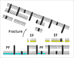
  • P-Face (or protoplasmic face; cyan) - most proteins (or complexes of proteins) remain embedded in the inner half of the plasma membrane adjacent to the cytoplasm. The P-face of the bottom plasma membrane was exposed by the fracture.
  • E-Face (or extracellular face; yellow) - fewer particles are found in the outer half of the plasma membrane adjacent to the extracellular space. The E-face of the top plasma membrane was exposed by the fracture.

Note: It is not possible to view both the P- and E-faces of one membrane in the same specimen.

© 2005-2026. T. Clark Brelje and Robert L. Sorenson